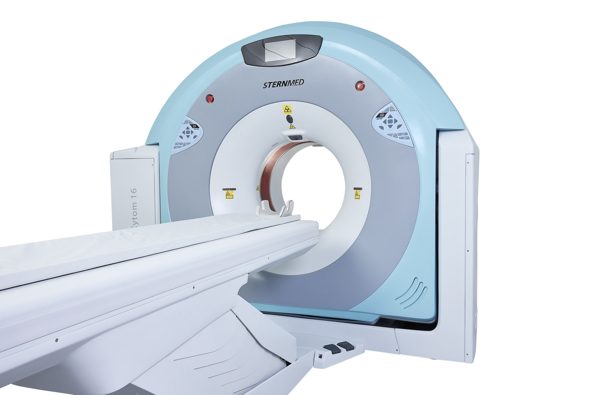
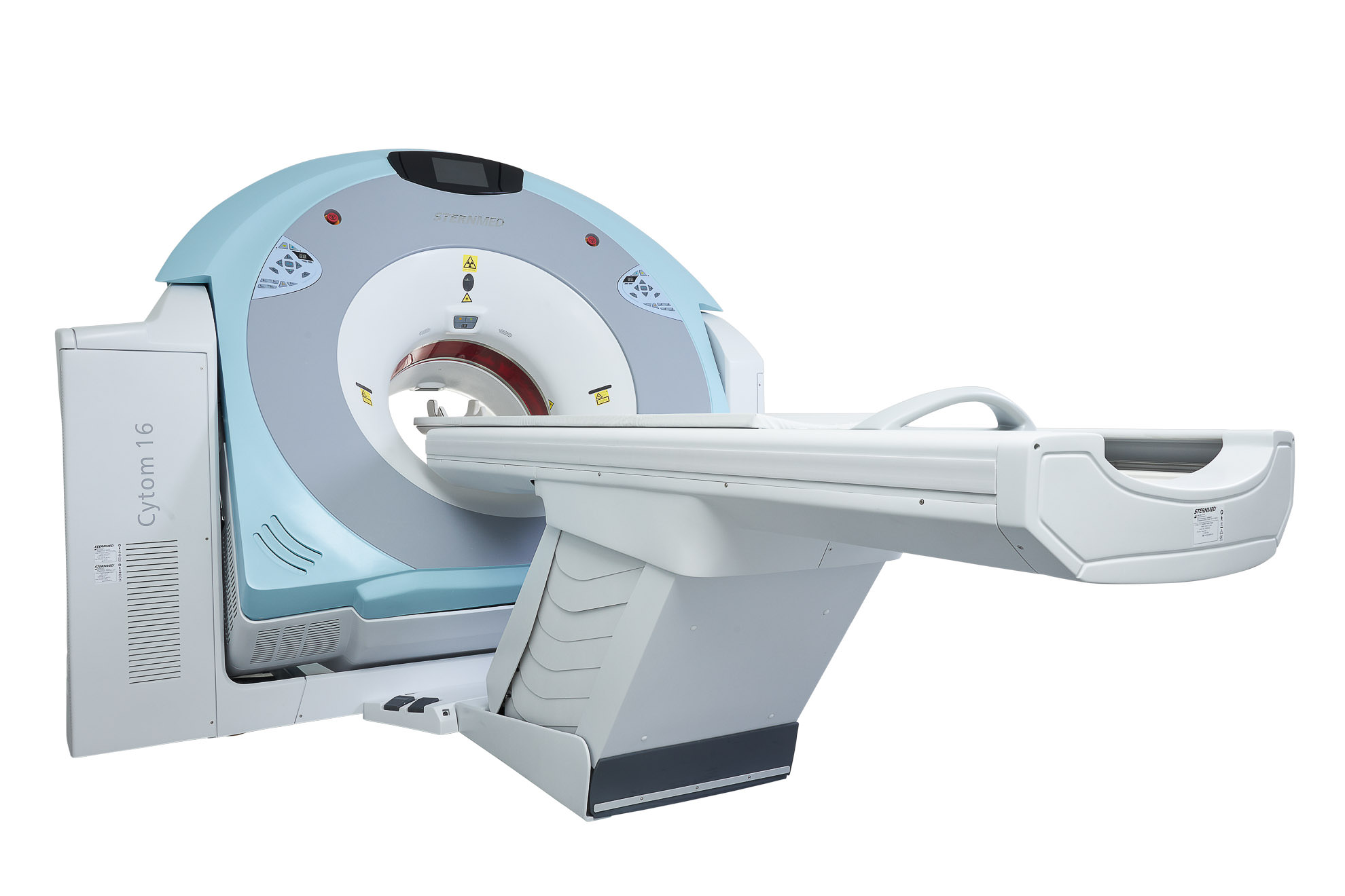
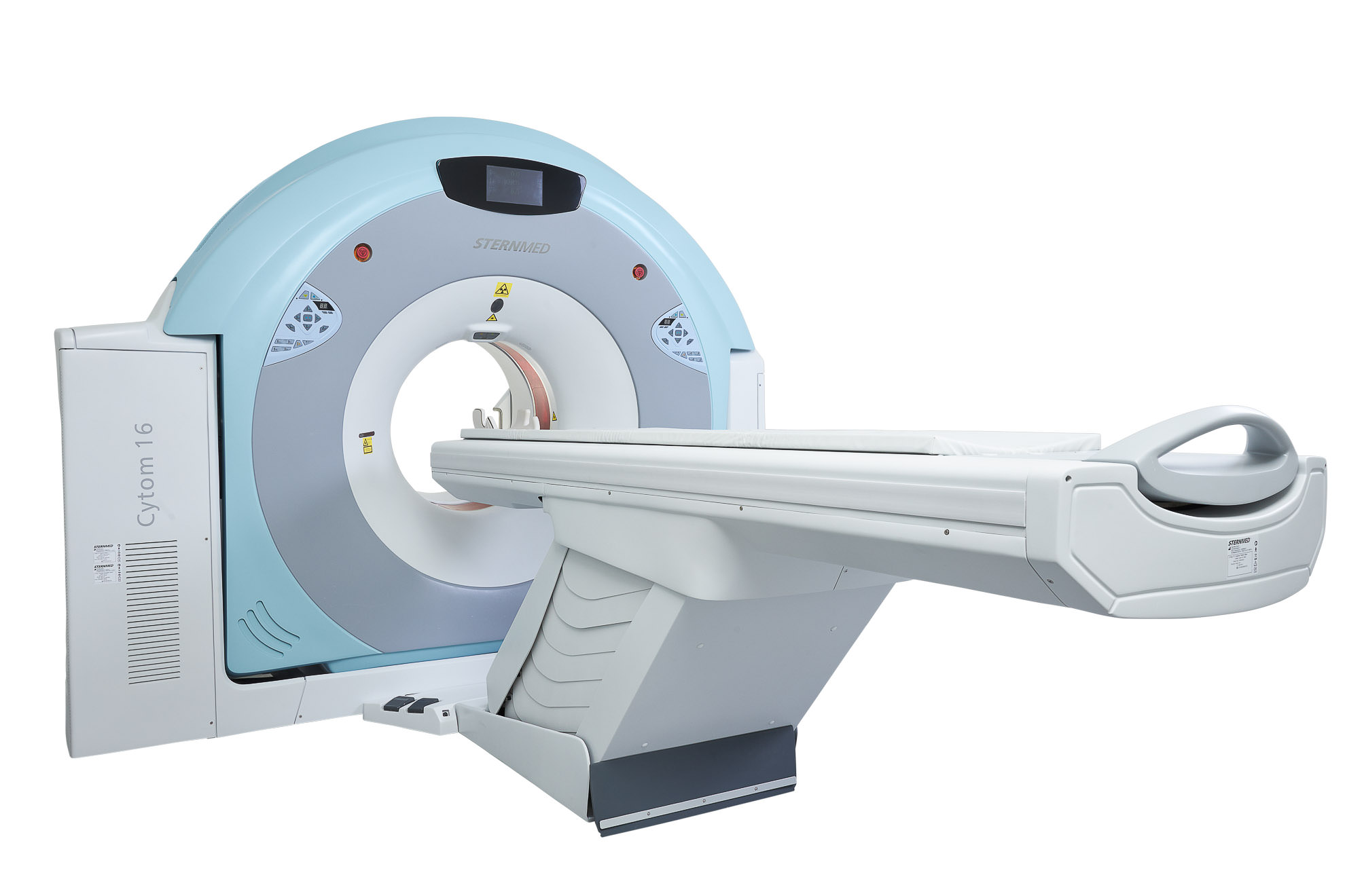
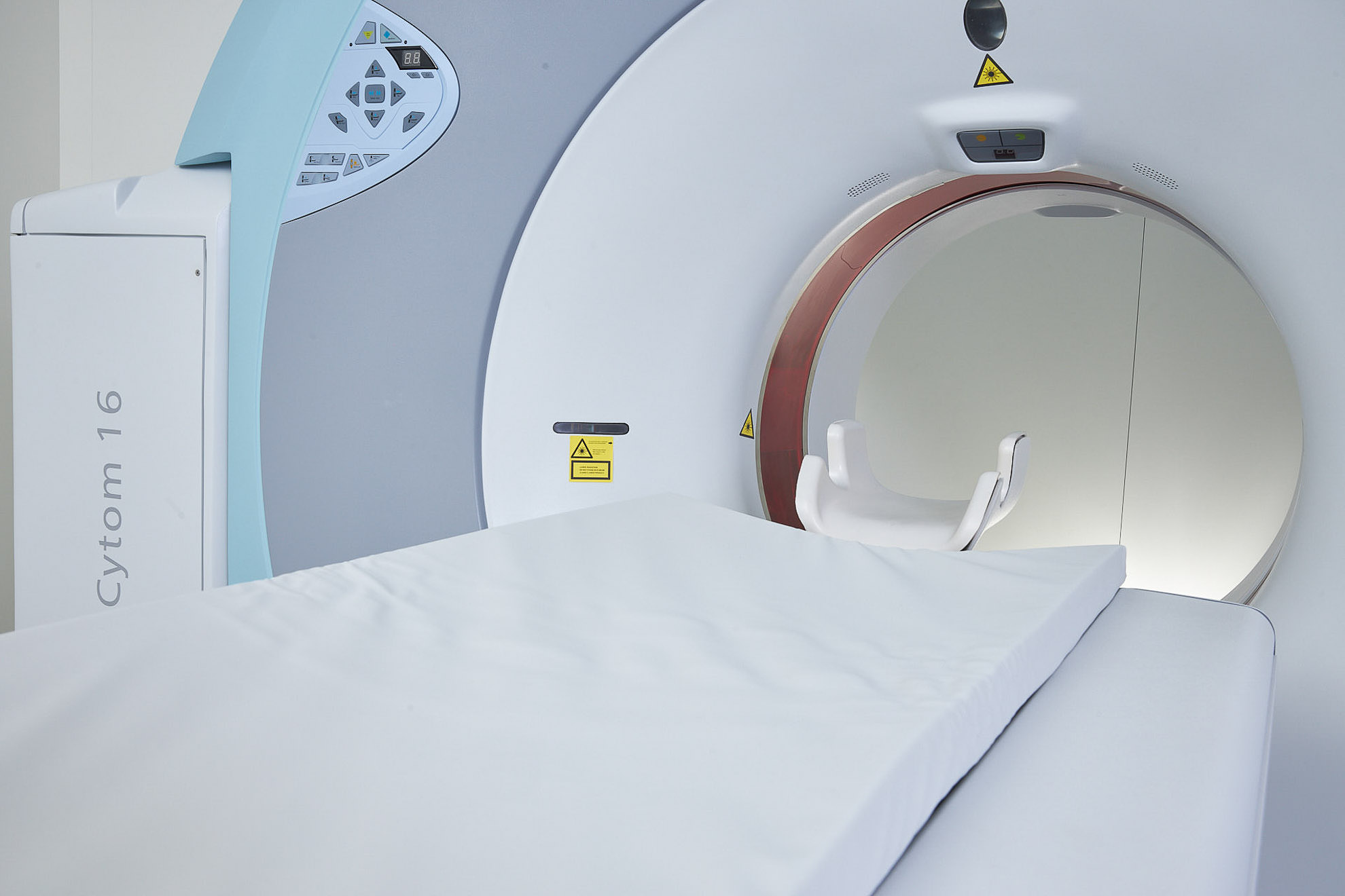
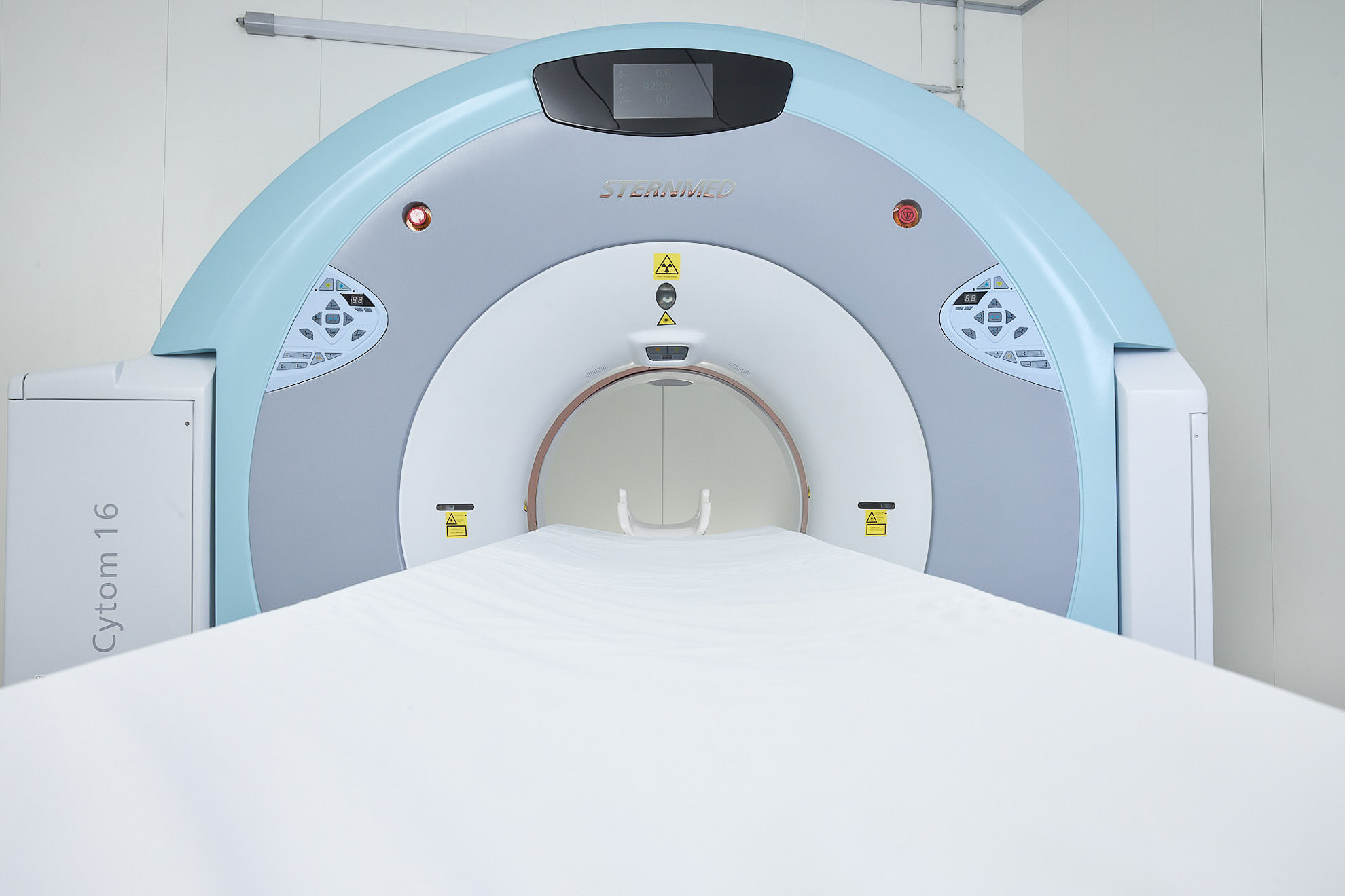
Cytom 16
Multi-slice ultra-fast CT scanner
Cytom 16 is a multi-slice ultra-fast CT scanner with PowerLink Non-Contact Power Technology. PowerLink eliminates the limitations of today’s rotating gantry systems providing the lowest cost, highest reliability CT power system, and non-contact slip ring design.
Features
World Leading Gantry Technology
- Higher Rotation Speed
- Faster Dynamic Response
- Smaller in Dimension & Weight
Highly Integrated Data Acquisition System
- Number of Detectors per Row 896
- Number of Detector Units 21504
- Minimum Slice Thickness 0.625mm
Perfect X-ray Generator System
- Power of Generator 60kW
- Tube kVp Range 80-140kV
- Tube mA Range 10-500mA
Non-contact Slip-ring Designed Innovative PowerLink™ Gantry Technology
- Never attrited and non-contact slip ring design
- Full integration in data communication and transmission
- Integrated control in gantry rotation
Advantage of Automatic dose control
- Tube current is automatically controlled according to the features of organs to reduce the radiation dose effectively on the basis of ensuring images quality.
- It is the latest and advanced technology for reducing dose, which can ensure high clinical image quality with large does reduction to the patient, even under the condition of 60%~70% normal dose.
- Pediatrics exclusive scanning solutions provides precise scan protocol for children, it offers dedicated scan parameters according to physical characteristics of children to reduce the radiation dose.
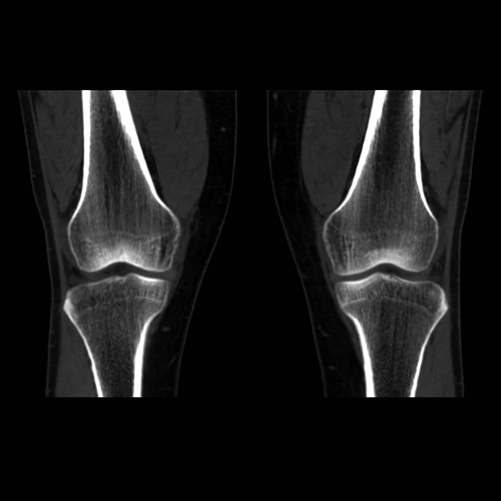
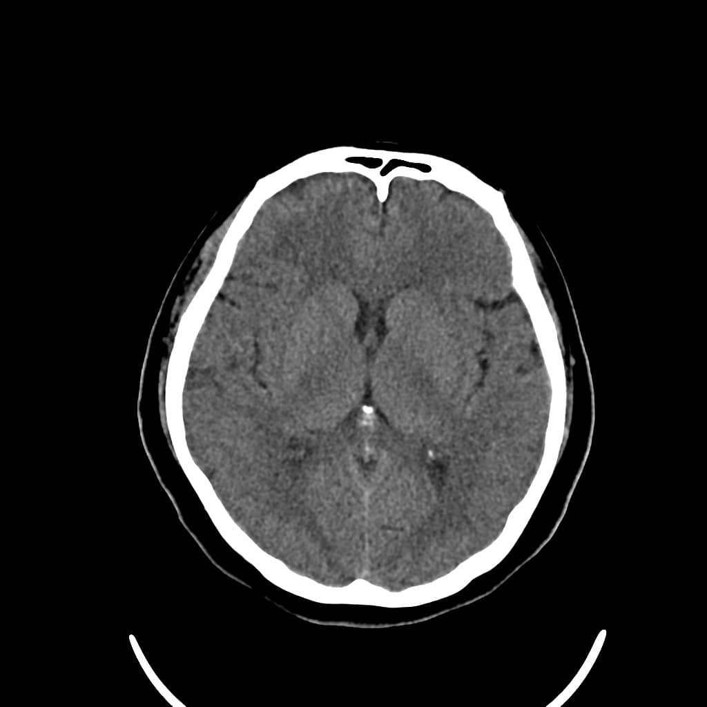
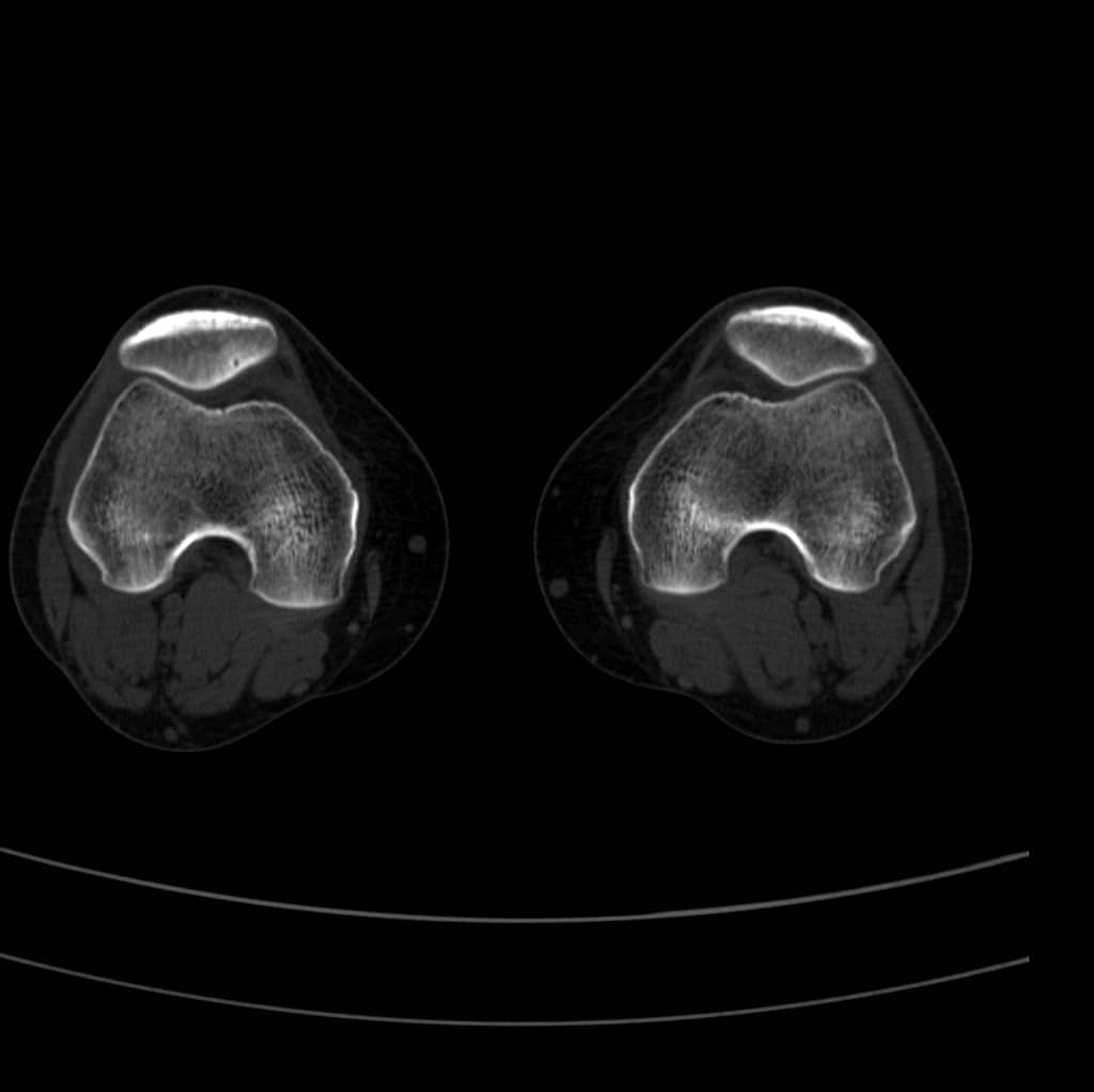
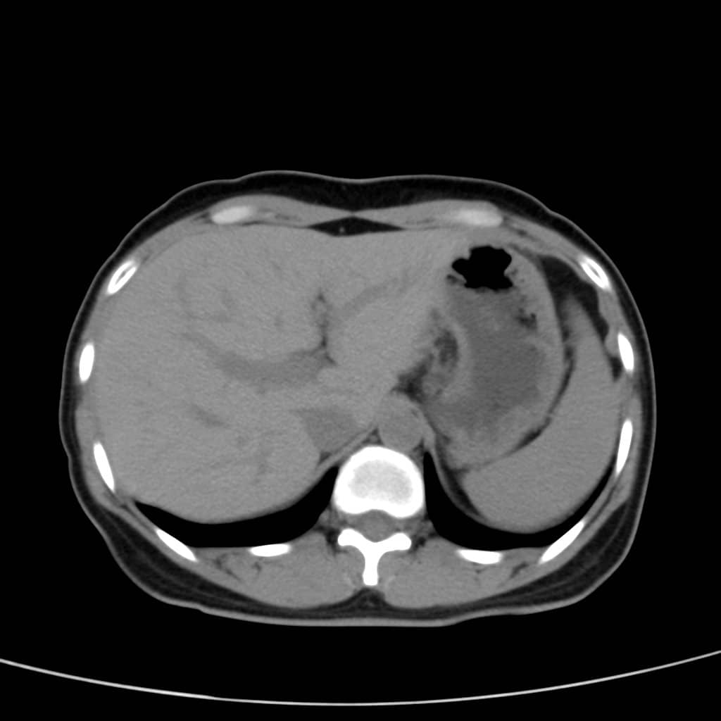
Outstanding Clinical Images
- Head: Conventional, Paranasal Sinus, MPR, SSD, VR, CTA, HD Inner Ear Cervical Vertebra: Conventional, MPR, VR, CTA
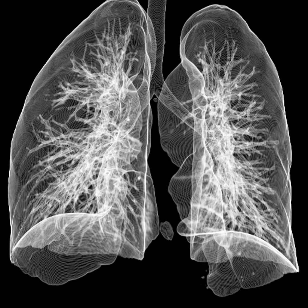
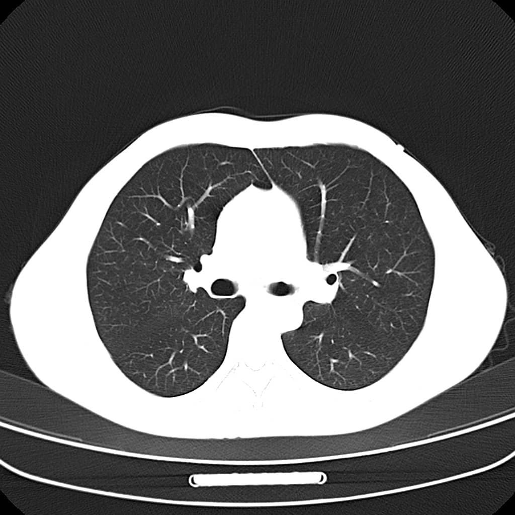
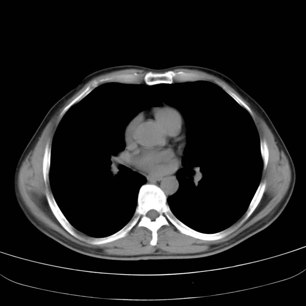
- Chest: Conventional (Lung Window, Mediastinal Window), MPR, VR, MinIP
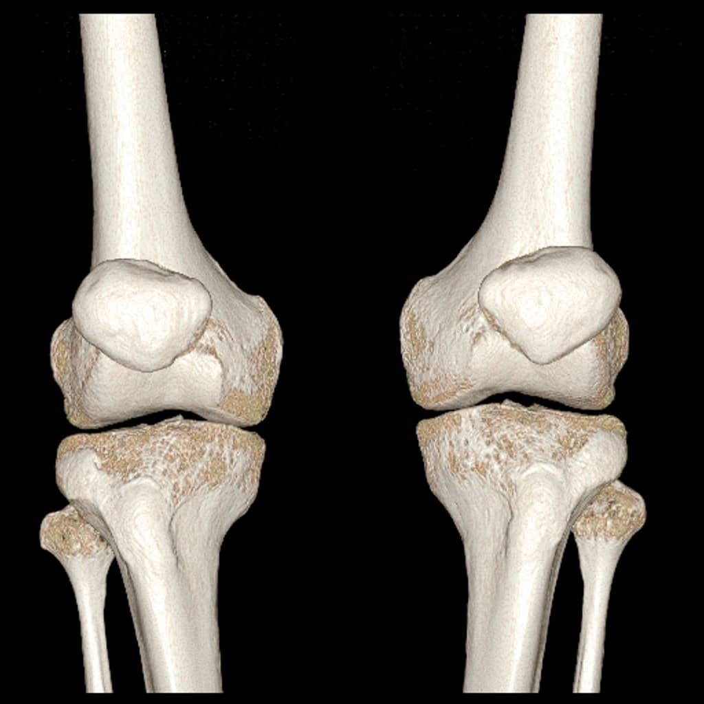
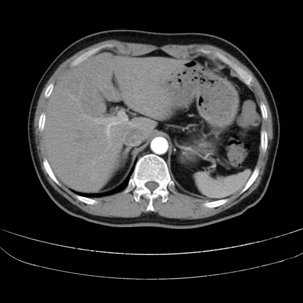
- Abdomen: Conventional, Enhancements Phases), MPR, VR, CTA Extremities and Joints: Conventional, MPR, VR
Minimum Maintenance Costs
- Lower dose, lower consumption
- Non-contact power transfer, without failures resulting from the brush slip-ring designs.
- Reduced downtime
Specifications
- 16 slice Helical CT Scanner
- 0.5s/360°, fully satisfies clinical demand
- Ultra Fast rare earth ceramic detector
- 24 rows detector, one disc rotation collect 16 slices images
- 21504 detector units; minimum slice thickness is 0.625mm
- Coverage of each single scan is 20mm
- Multiple X-ray dose reduction technology
- Intelligent scattering line automatic elimination technology
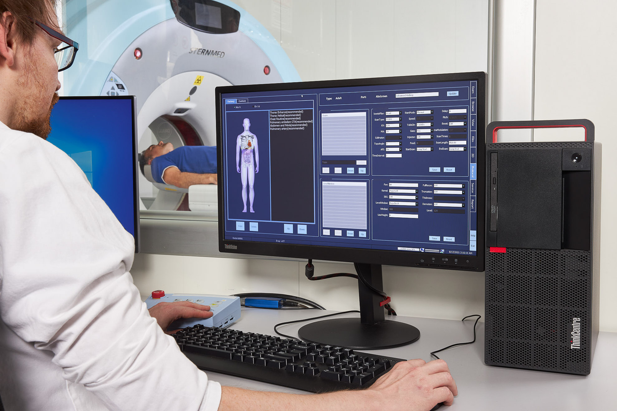
- Intelligent operation process
- High image quality
- Fast scan speed
- 120 Second non-Stop scanning
- Low x-ray dosage
Packages
3D image reconstruction Includes VRT, MPR, CPR, SSD, Simulated scalpel, Virtual endoscope, CTA Remove bone, CTA subtraction,etc.
Tissue Fluoro Combine 2D and 3D Image; Multi-Date field fusion for observation; Read a slice on any angles; Automatic push functions can reduce operation and improve speed
Tissue Element Analysis Make extraction and analysis of the tissue within the organ, giving quantitative analysis for the estimate of diagnose\treatment\treatment effect
CT Vessel Analysis After CTA subtraction or bone removal, make vessel straighten analysis, virtual endoscope, plaque analysis,virtual stent and so one, including head and neck, thoraco-abdominal, limbs and other parts.
CT Perfusion analysis According to the analysis of the dynamics of blood, detect the pathological field before the morphological changes occur. Multiple Perfusion Models including CT Head Model, CT Body Model, CT Liver Model; providing various parameters for analysis, including BF, BV, MTT, TTP, tMIP, PS, etc.
CT coronary artery analysis Mainly focus on cardiac CTA analysis, including One-key heart 3D reconstruction, panoramic view of heart,stenosis analysis, plaques analysis, vessel straighten analysis, broken branch and CTO reconstruction, Heart display as a globe, virtual stents placement and measurement Automatic analysis and manual correction are both available.
CT Calcium score Analysis According to calcium score, evaluate the quality of the coronary and give quantitative analysis of the calcium volume, CT value to assist diagnosis.
CT Heart Function Evaluation Dynamic play and AVI storage of the cardiac motion; automatically calculate the parameters of the cardiac function, such as Ejection Fraction, End.Sys.Vol, End.Dia.Vol, ventricular wall movement, ventricular wall thickness, etc.
CT Bone mineral density analysis Calculate and display the bone mineral density of the ROI. TScore and ZScore can be automatically calculated or self defined analysis. Mineral density analysis report is created by one key. Images of mineral density measurement and analysis are saved as DICOM.
CT spine extraction and analysis Whole spine is segmented automatically. Offer Segmental analysis, auto-name, virtual multi-dimensional X-ray images, one-key multiple slices printing, etc. Spine diagnosis becomes quite efficient.
CT rib analysis Auto-analysis of the rib. One-key segmentation of the target rib, display 3D view and CPR. Full analysis of the rib makes the diagnosis quite efficient.
CT Colon Analysis Forward path and backward path of virtual colonoscope, self-navigation, colon unfolding display to help doctor to observe the inner side. Auto segmentation of analysis of the interested colon segment and mark the lesion.Various measurement parameters for the lesion are provided.
CT Lung nubble Analysis Analysis of the suspected lesion within the lung. Automatic lung extraction and manual extraction are both available. Nubble volume measurement, evaluate the nubble size changes through time, assist to qualitatively determine the nubble. In addition, various parameters are provided, including volume, CT value, component analysis, curves, etc.
CT pulmonary edema analysis Calculate the volume of ROI, water volume, water weight, density in the lung CT image, display the ROI analysis result in the list. Assist the pulmonary edema diagnosis.
CT Lung analysis One-key to segment the airway and left/right lung with pseudo color processing;Quantitative and qualitative analysis of the lung.
CT lung markings analysis Auto-segmentation of the whole lung;For the lung nubble, generate the voxel analysis curve and data form.
Advanced Dental Radiological Analysis function Dental full view and sectional view, provide various measurements, assist dental diagnosis and to make operation plans.
CTU Urological imaging analysis
CT liver Analysis Auxiliary to develop operation plan based on CT images; image processing includes liver segment, extracting blood vessels, liver surgery simulation
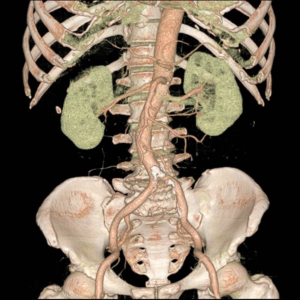
CT Abdominal panoramic reconstruction Based on contrast-enhanced CT abdomen images, offers 3D reconstruction, tissue extraction with the fusion display, reconstruction of the abdomen panorama, better diagnosis and operation programming.
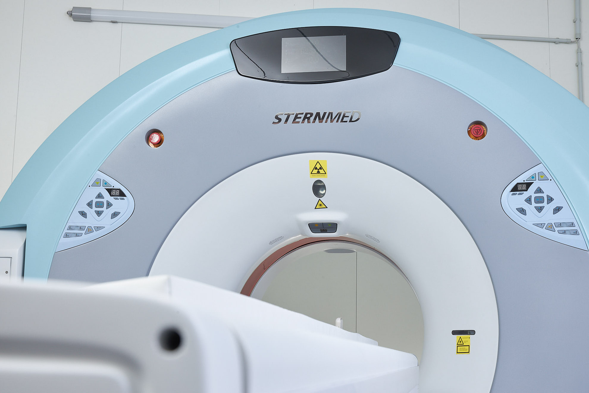
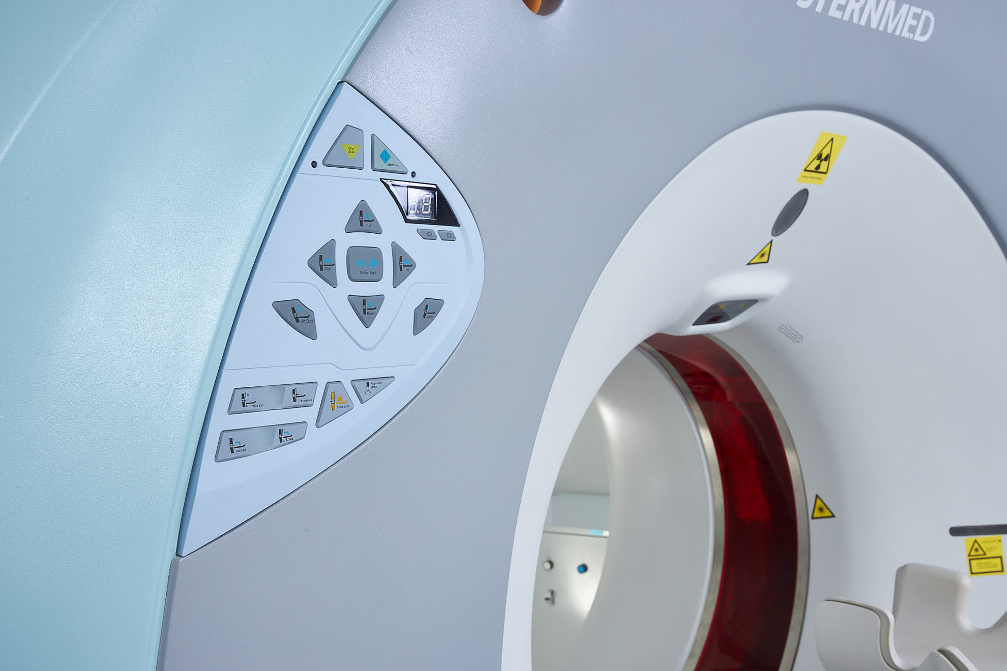
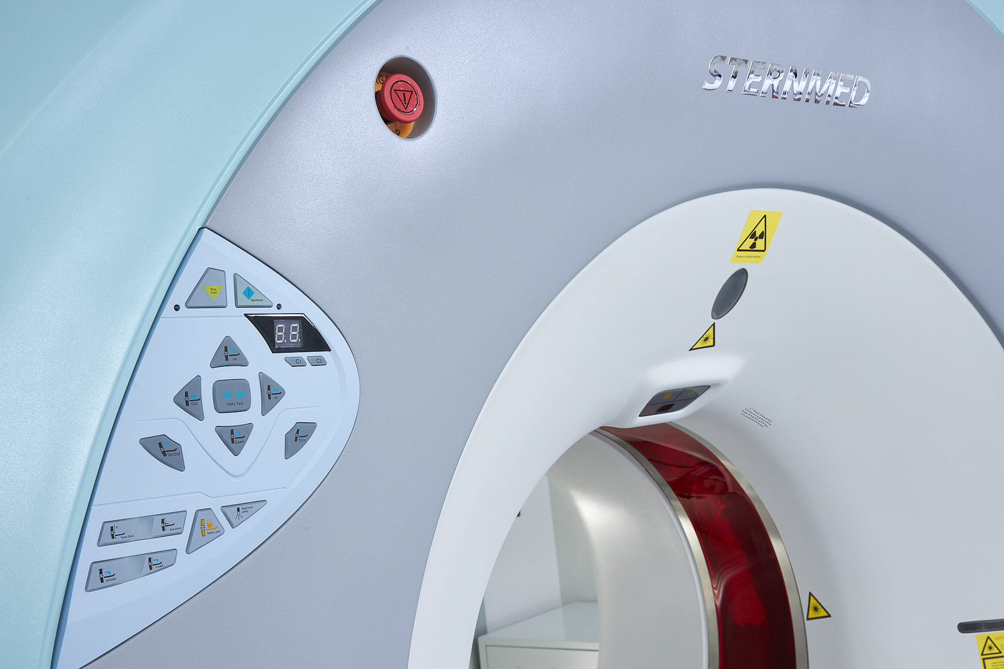
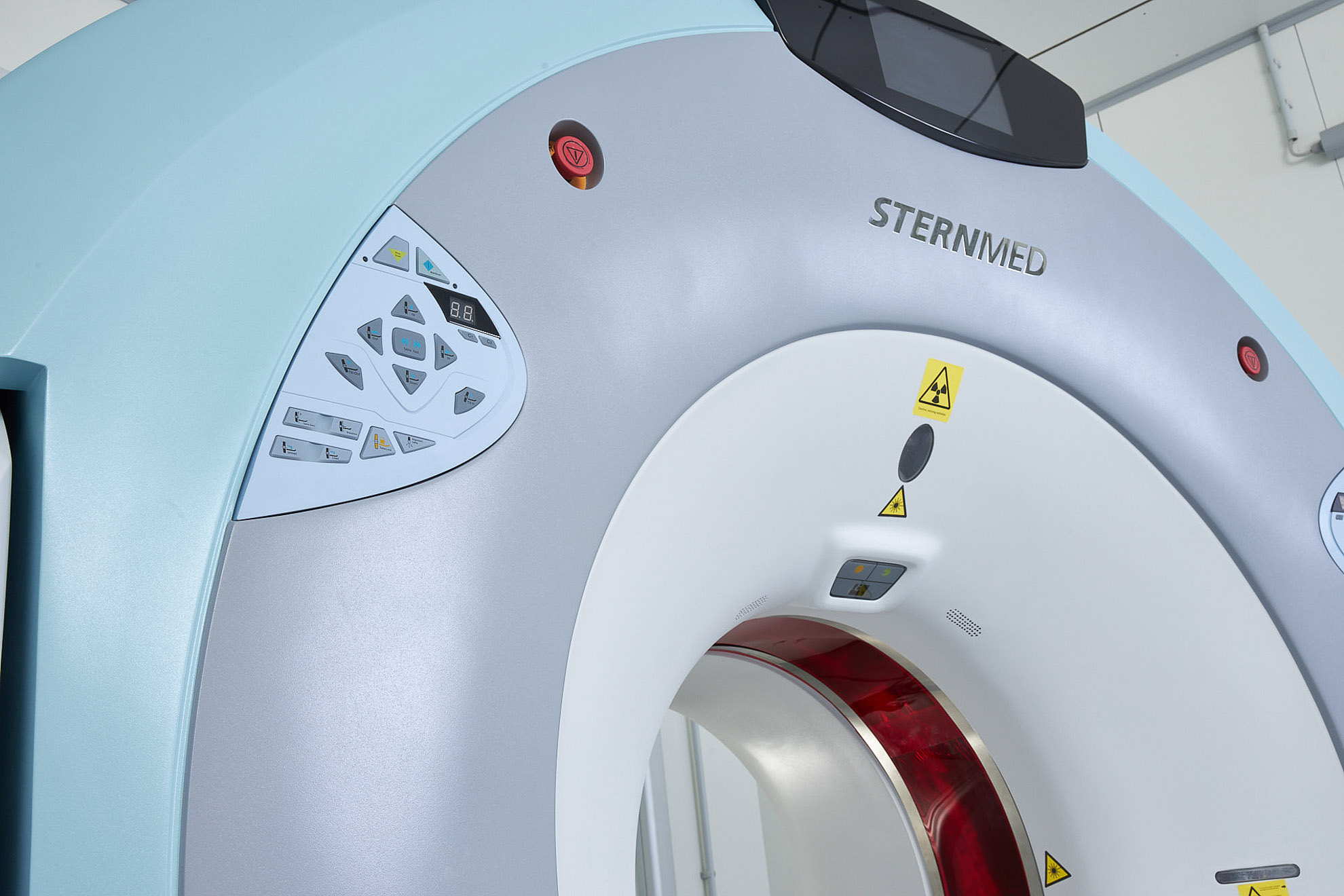
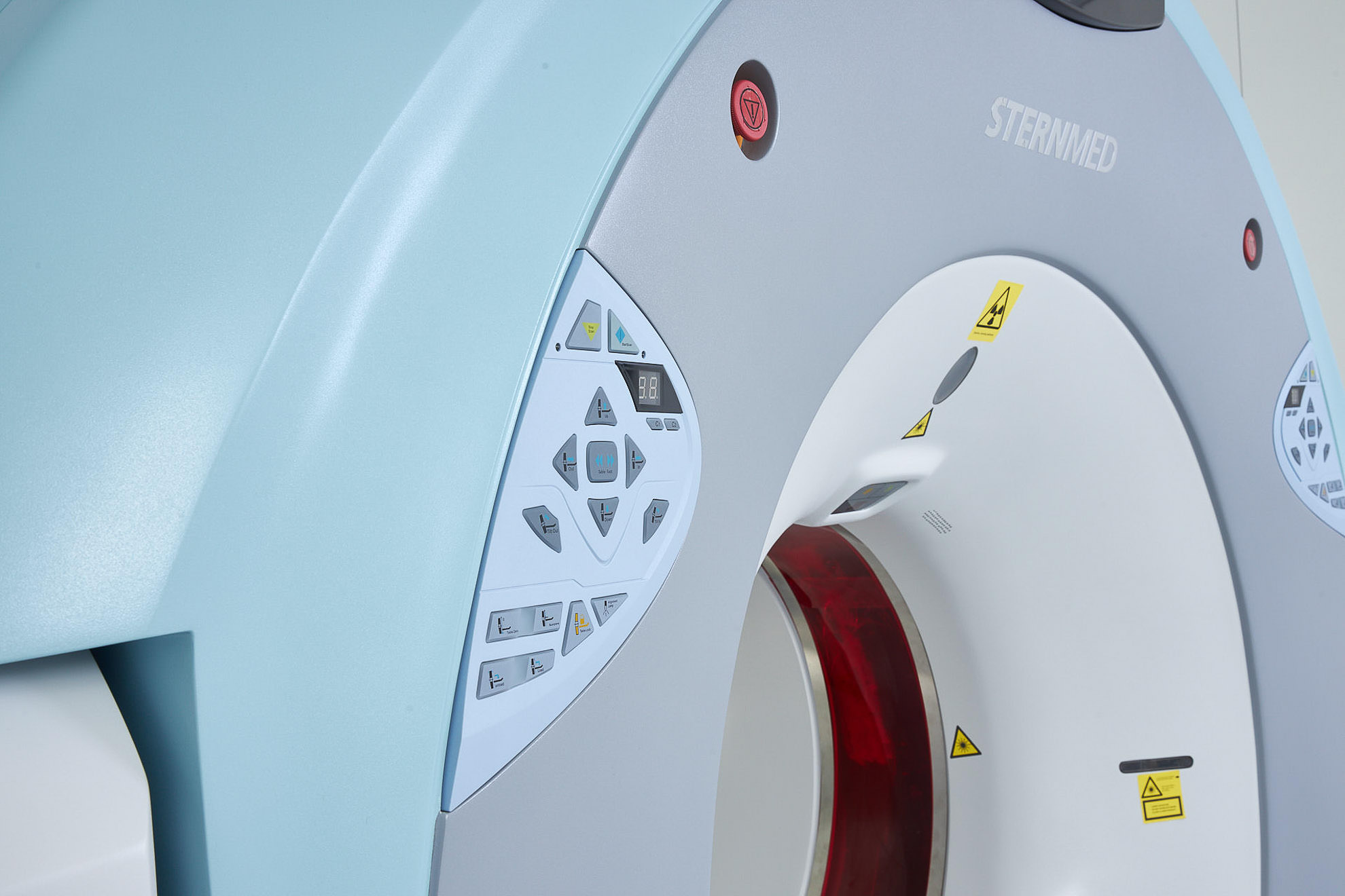
Product images
Clinical Use / Images